


A Peek into the First Days of Human Life
High-Resolution Imaging of Developing Embryos

Imagine if we could watch a tiny human life grow from the very beginning, from just a few cells to a fully formed baby. Well, scientists have just taken a big step towards making this a reality. They have managed to capture the most detailed images ever of human embryos growing in real time. And the best part? They did it without having to alter the embryos in any way, which is a big deal because it avoids ethical issues.

So, how did they do it? The researchers used two common laboratory tools: fluorescent dyes and laser microscopes. The fluorescent dyes are special markers that can be added to a sample to highlight certain cell structures. The laser microscopes are like super-powered magnifying glasses that can see tiny details in the embryos.
The embryos used in this study were donated by an IVF clinic and were at a very early stage of development, with only 60–100 cells each. The researchers used two types of fluorescent dyes, one that labels DNA and another that labels a protein that forms the skeleton of cells. They then watched the embryos develop over the first 40 hours using the laser microscopes.
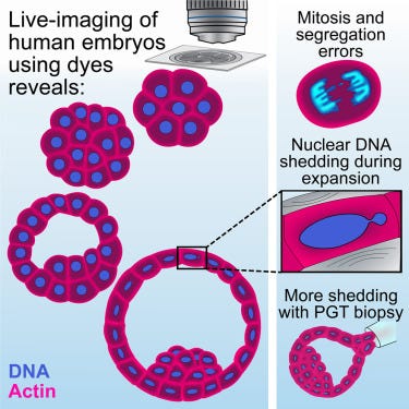
What they saw was amazing. They could see the cells dividing and the chromosomes separating. They even noticed some cells losing some of their DNA during a stage of cell replication. This could be linked to chromosomal abnormalities that can cause pregnancy loss and failure of implantation.
The researchers also compared human embryos to mouse embryos, which are often used as models to study embryonic development. They found some important differences, like the timing and process of cell shape changes. These small differences could have big impacts on how the uterus develops.
This new imaging technique could lead to the development of ways to non-invasively screen embryos conceived through in vitro fertilization (IVF). This means doctors could use this type of live-imaging to follow embryos in the clinic and determine which embryo is likely to have the best potential before implantation.
But this is just the beginning. The researchers hope to build on this research by imaging human embryos for longer periods, using lower-intensity laser microscopes, and incorporating other dyes that can label different structures, such as cell membranes.
This breakthrough is like having a new set of eyes to see the earliest stages of human life. It's a big step forward in understanding how we develop and could lead to new ways to help ensure healthy pregnancies. So, the next time you see a baby, remember that they started as just a few cells, and scientists are working hard to understand that magical journey.
References and image credits:
doi: https://doi.org/10.1038/d41586-023-02222-3
Domingo-Muelas, A. et al. Cell https://doi.org/10.1016/j.cell.2023.06.003 (2023).